Our Services
The Lump and Bump Doc practice was created to provide a novel approach towards the diagnosis and management of skin lesions, particularly 'lumps and bumps'.
1.
Skin Cysts
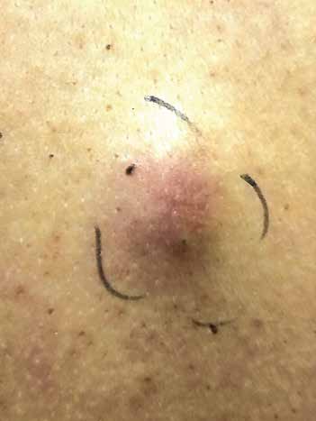
Before
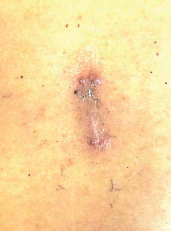
After
These are usually put under the general designation of “lumps and bumps” and are extremely common in everyday practice. Cysts are cavities that are lined by epithelium. Cysts come in various forms and dimensions and cover a broad swath of potential common skin lesions. Some of the common forms or names are pilar (aka trichilemmal), epidermoid, infundibular, dermoid and sebaceous.
Often patients present with a cyst that’s acutely inflamed. In those circumstances it's usually prudent to treat the underlying inflammation before attempting excision, increasing the chances for improved wound healing. The usual indications for cyst removal are: Rapid growth, increasing pain, drainage, unsightly appearance or uncertain etiology. This procedure termed a ‘cystectomy’ is performed in the office under local anesthesia.
2.
Lipomas
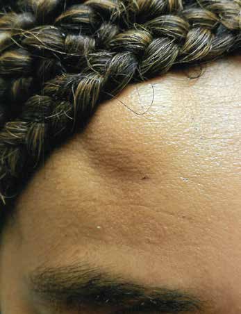
Before
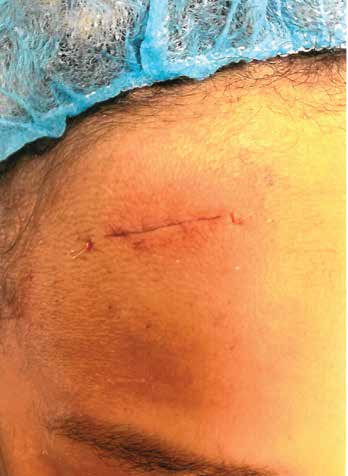
After
Lipomas are growths usually under the skin (subcutaneous) and are composed of lobules of ‘fatty tissue’ that’s surrounded by a capsule. They’re usually benign but externally it is difficult to know if the growth is just fatty tissue or some other type of more serious lesion. In those circumstances, or if there’s rapid growth, excision is warranted. Other situations necessitating excision are if there’s a suspicion or uncertainty as to the pathology or if the lesions’ location is unsightly or causes discomfort. These lesions are treated commonly in the office by excision using local anesthesia.
3.
Moles (aka Nevi)
Before
After
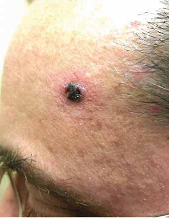
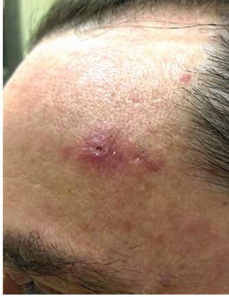
Nevi or as they’re commonly called “moles” are flat or slightly raised skin growths that are usually hyperpigmented. They can appear or grow anywhere on the body’s surface. They’re usually non-painful, but can have smooth, rounded or irregular borders. They’re usually of uniform color but occasionally can be multi-colored with an irregular surface. Some moles can have a benign presentation but can also be a prelude to cancerous progression (melanocytic or dysplastic nevus) OR be suggestive of a malignant lesion such as malignant melanoma. In certain ethnic or racial groups there may be a tendency for these nevi to be or progress to more serious pathology and this necessitates greater surveillance, scrutiny and excision for histopathological examination. These lesions can be treated by either an incisional or an excisional biopsy under local anesthesia.
4.
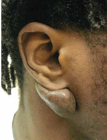
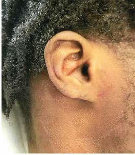
Keloids
Before
After
Keloids are manifested as excessive or overgrowth of healing tissue or overgrowth of a scar (hypertrophic scar). The underlying pathology is excessive or out of control collagen growth or proliferation. They can be small and barely perceptible or extremely large in areas such as the ear lobe (where the lobe was pierced for an earring)), the arm (at an injection site), or at a surgical incision site.
There are currently several modes of treatment, including injection with steroids, pressure and massage, injection with immunomodulating meds, cryosurgery (freezing), radiation or, excision (i.e., surgical removal). Unfortunately, the recurrence rate for keloids after any treatment is relatively high.
5.
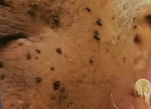
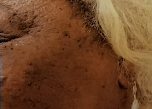
Skin Tags (Acrochordons)
Before
After
These are very common ‘fleshy’ skin lesions most notable on the face and neck. They’re usually dark in color, but rarely painful, but can be in areas that cause them to rub or irritate by clothing, necklaces or eyewear. They’re treated by clipping or cautery with a small amount of local anesthesia. They’re usually treated or removed in groups of 5.
6.
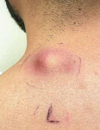
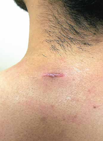
Skin Abscesses
Before
After
A skin abscess is a bump that appears within or below the skin’s surface. This bump is usually full of pus or translucent fluid. It’s typically due to a bacterial infection. Most skin abscesses are harmless and may go away without treatment. Sometimes, skin abscesses are more difficult to treat and may require Incision & Drainage ( I&D ).
Common causes include bacteria due to poor hygiene, chronic skin disease, a weakened immune system, or even infected hair follicles.
7.
Lab Services
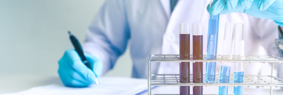
As an adjunctive arm of our derm practice we offer lab services through an entity called Lab Tests To Go. This service is unique and is available to walk-in customers as well as established patients who can have labs drawn at our office the same day or by appointment that they (or their physicians’ request) request without a physician’s order.
We offer a full range of lab studies, including but not limited to: CBC, CMP, BMP, Liver function studies, Kidney studies, Cholesterol, PSA, A1c (for those with diabetes), STD tests, and Urine drug screens (15-minute Rapid in-office or sent out to an Accredited lab). Payment works the same as with our derm skin surgery practice: Payment is at time of service; No insurance is necessary or prior approval.